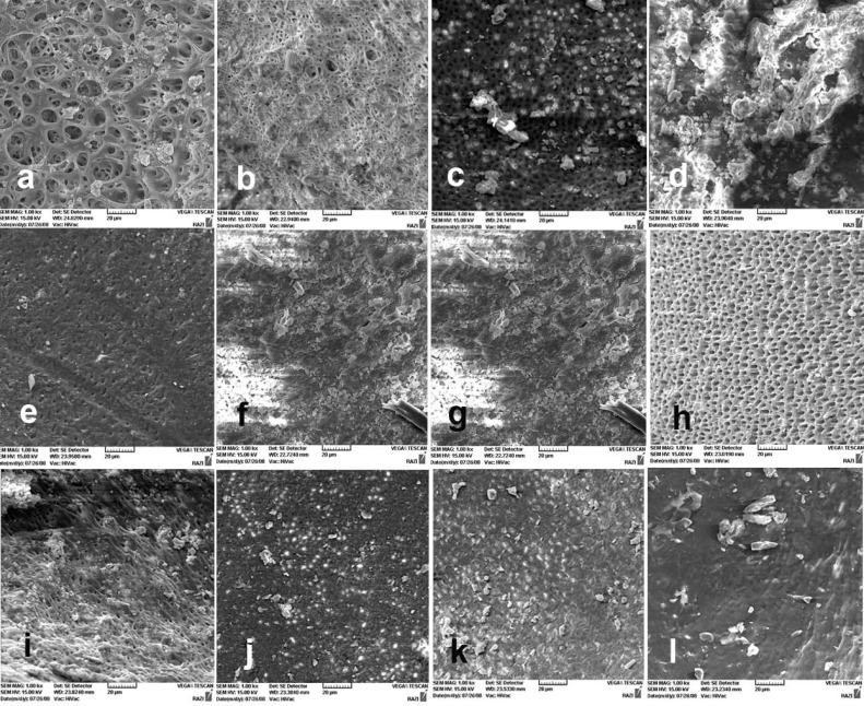
Figure 1.
A) The coronal third of the canal wall after using 1% papain solution; almost no smear layer is remained. Orifice of dentinal tubules are patent (score 1); B) The middle third of the canal wall after using 1% papain solution (score 1); C) The apical third of the canal wall after using 1% papain solution, small amount of smear layer, some open dentinal tubules are visible (score 2); D) The coronal third of the prepared canal wall after using 0.1% papain solution; note the homogenous smear layer along almost the entire canal wall. Only very few open dentinal tubules are present (score 3); E) The middle third of the prepared canal wall after using 0.1% papain solution; the entire root canal wall is covered with a homogenous smear layer and open dentinal tubules are absent (score 4); F) The apical third of the prepared canal wall after using 0.1% papain solution; a thick, homogenous smear layer covering the entire root canal wall is evident (score 5). The canal wall in negative control sample; G) The clean canal wall in the coronal third of the prepared canal (score 1); H) The clean canal wall in the middle third of the prepared canal (score 1); and I) The canal wall in the apical third (score 2). The canal wall in positive control sample; J) The coronal third of the prepared canal (score 5); K) The middle third of the prepared canal (score 4); and L) The apical third of the prepared canal (score 5)

