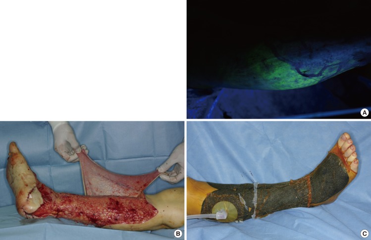
Fig. 1.
Intraoperative photographs demonstrate the authors' methods
(A) Under Wood's lamp illumination areas of fluorescence and non-fluorescence and mottled areas can be distinguished. (B) The flap of the non-fluorescent area is defatted to be used as FTSG. (C) To prevent hematoma formation under FTSG, VAC dressing is applied. FTSG, full-thickness skin graft; VAC, vacuum-assisted closure.

