
FIGURE 2.
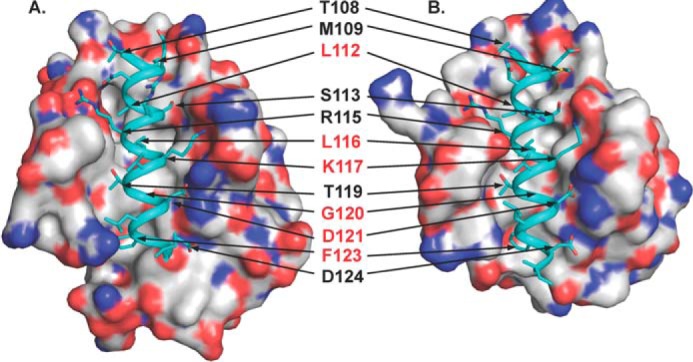
Comparison of the WT Beclin 1 BH3 domain bound to Bcl-XL (Protein Data Bank code 2PIL, A) and M11 (Protein Data Bank code 3DVU, B). Each complex is shown in a superimposable view with Bcl-XL and M11 shown as molecular surface colored by atom type: oxygen, red; nitrogen, blue; sulfur, yellow; and carbon, light gray. In each complex structure, the WT BH3 domain is rendered as teal ribbon, and residues are displayed in stick. The 12 BH3 domain residues involved in binding to both Bcl-XL and M11 are labeled, with the residues selected for mutagenesis highlighted in red. This and all other molecular figures were prepared with the program PyMOL.