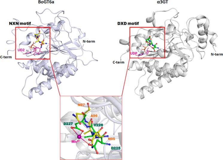
FIGURE 1.
Structural comparison of the metal independent BoGT6a (with the NXN motif in silver) and metal dependent bovine α3GT (with the DXD motif in gray). The inset shows the details of the NXN motif (in yellow) and DXD (in green) motifs in BoGT6a (12) and α3GT (Protein Data Bank (PDB) code 1K4V (17)), respectively. The bound Mn2+ ion in α3GT is shown in purple sphere.

