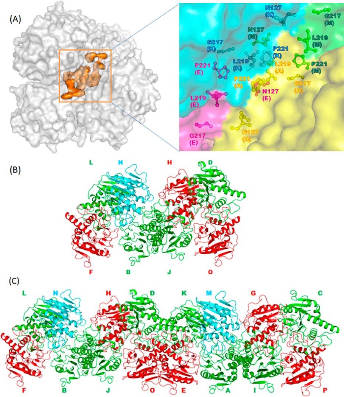
FIGURE 4.
Arrangements and interactions of BoGT6a molecules. A, packing between chains in the monoclinic form III structure showing hydrophobic core interactions. The surfaces of protein molecules are colored gray, and the hydrophobic core is colored orange, respectively. The inset shows the details of the hydrophobic core in which the hydrophobic residues are displayed as sticks and colored by chains. B, arrangement of the minimal packing unit in the monoclinic crystal form. Structure A is in green; structure B is in red, structure C is in cyan, and protein molecules are shown as graphics. C, arrangement of 16 molecules in the asymmetric unit in monoclinic crystal form.

