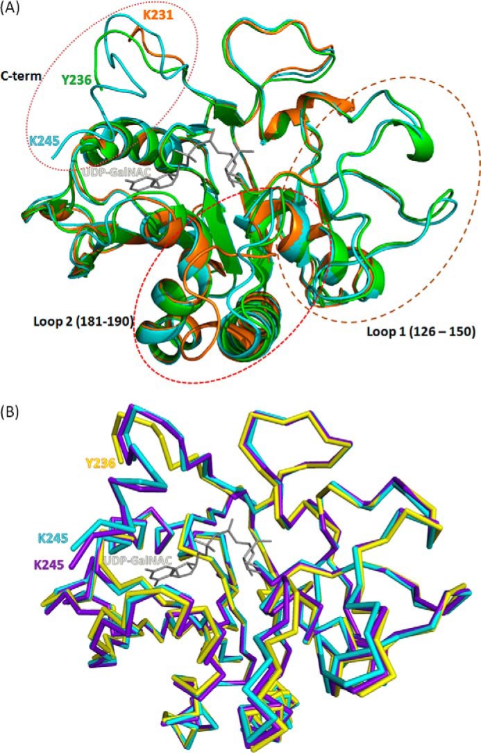
FIGURE 5.

Effects of binding different ligands on the conformation of BoGT6a. A, superimposed ribbon structures of apo-protein (orange), FAL complex (green), and UDP-GalNAc complex (cyan). The UDP-GalNAc ligand from the latter complex is colored gray. The marked loops are regions that undergo major structural adjustments and are discussed under “Results and Discussion.” B, superimposed ribbon structures of chain A (structure B, yellow), chain E (structure A, cyan), and chain M (structure C, purple). The UDP-GalNAc ligand from chain E is colored gray.
