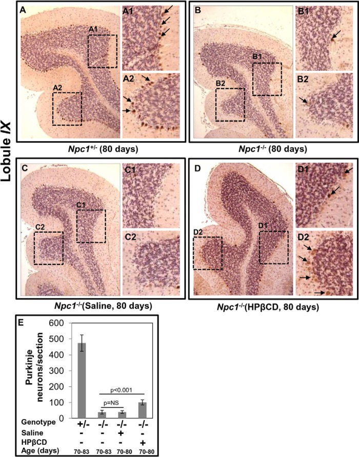
FIGURE 6.
Effects of cyclodextrin on the Npc1nih mouse brain as determined by immunohistochemistry. Formalin-fixed paraffin-embedded brains were sectioned (sagittally, 4–5 μm) and stained using anti-mouse calbindin antibodies to visualize Purkinje neurons in the entire cerebellum. Micrographs shown are representative images of the IX lobule of the cerebellum. A, Purkinje neurons (stained in brown) indicated by black arrows are evident in Npc1+/− mice (age, 80 days). A1 and A2 are the magnified areas boxed in A. B, loss of Purkinje cells in the cerebellum of an Npc1−/− mouse (age, 80 days). Calbindin immunoreactivity was barely detected across the different lobules of cerebellum. B1 and B2 are magnified areas boxed in B. C, cerebellar section of an Npc1−/− mouse (age, 80 days) injected with saline was devoid of Purkinje neurons. C1 and C2 are magnified areas boxed in C. D, chronic HPβCD treatment (that partially rescued inflammation) also partially recovered Purkinje neurons in Npc1−/− mice. A few lightly brown stained Purkinje neurons (indicated by arrows) are seen. D1 and D2 are magnified areas boxed in D. Original magnifications, ×40. E, semiquantitative analysis of Purkinje neurons in an Npc1nih mouse brain. Numbers of Purkinje neurons in the calbindin-labeled cerebellar sections from four mice (age, 80 days) in each group were counted. HPβCD treatment resulted in a small but significant increase in the number of Purkinje neurons. The data represent the mean ± S.D. (error bars). NS, not significant.

