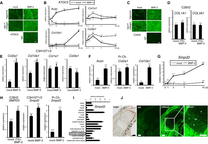
FIGURE 1.
Expression of Smpd3/nSMase2 is promoted by BMP-2 treatment in maturing chondrocytes in vitro and is increased in prehypertrophic and hypertrophic chondrocytes in a growth plate in vivo. A, chondrogenic differentiation of mouse ATDC5 chondrocytes was induced by application of BMP-2 (300 ng/ml) for 7 days (d). Immunofluorescence for aggrecan or type II collagen was performed, with normal IgG as negative control. Scale bar, 100 μm. B, ATDC5 cells were cultured in the presence of BMP-2 (300 ng/ml) for the indicated periods. Expression levels of Acan, Col10a1, Col1a1, and Col3a1 were examined by quantitative RT-PCR. C, chondrogenic differentiation of human C28/I2 chondrocytes was induced by the application of BMP-2 (300 ng/ml) for 7 days. Immunocytochemistry for aggrecan or type II collagen was performed, with normal IgG as negative control. Scale bar, 100 μm. D, C28/I2 chondrocytes were cultured in presence of BMP-2 (300 ng/ml) and ITS supplement for 14 days. Expression of COL1A1 or COL3A1 was evaluated by quantitative RT-PCR. E, mouse C3H10T1/2 cells were cultured with BMP-2 (300 ng/ml) for 48 h. Expression levels of Col2a1, Col10a1, Col1a1, and Col3a1 were evaluated by quantitative RT-PCR. F, mouse primary chondrocytes (Pr.Ch.) cells were cultured in the presence of BMP-2 (300 ng/ml) for 6 days. Expression levels of Acan, Col2a1, and Col10a1 were evaluated by quantitative RT-PCR. G, ATDC5 cells were cultured in presence of BMP-2 (300 ng/ml) for the indicated periods. Expression of Smpd3 was examined by quantitative RT-PCR. H, quantitative RT-PCR for Smpd3 was performed on samples in D, E, and F. I, real time PCR for Smpd3 was performed on a tissue cDNA panel of a 3-month-old mouse. J, expression of nSMase2 in mouse E17.5 humerus cartilage was evaluated by immunofluorescence. Normal IgG was used as negative control. r, resting chondrocytes; p, proliferating chondrocytes; ph, prehypertrophic chondrocytes; h, hypertrophic chondrocytes; b, bone. Scale bar, 200 μm. *, p < 0.05; **, p < 0.01. ΔCt values (Ct target − Ct Hprt1) of quantitative RT-PCR are indicated in the graphs.

