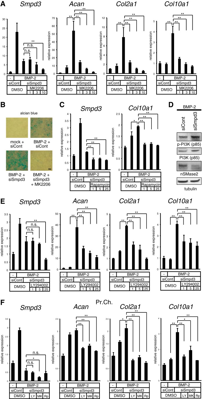
FIGURE 5.
Blocking the Akt or PI3K pathway negates the Smpd3 siRNA-mediated acceleration of chondrogenesis initiated by BMP-2 in ATDC5 cells. A, ATDC5 cells were transfected with control siRNA (siCont) or Smpd3 siRNA (siSmpd3) for 16 h and stimulated by BMP-2 (300 ng/ml) with or without MK2206 at the indicated concentrations (micromolar) for 6 days. Expression of Smpd3, Acan, Col2a1, and Col10a1 was evaluated by quantitative RT-PCR. B, ATDC5 cells were transfected with control siRNA (siCont) or Smpd3 siRNA (siSmpd3) for 16 h, and then cultured in the presence of BMP-2 (300 ng/ml) with or without MK2206 (10 μm) for 9 days. Alcian blue staining was performed. Scale bar, 300 μm. C, ATDC5 cells were transfected with control siRNA (siCont) or Smpd3 siRNA (siSmpd3) for 16 h and stimulated by BMP-2 (300 ng/ml) with or without rapamycin at the indicated concentrations (micromolar) for 3 days. Expression of Smpd3 and Col10a1 was evaluated by quantitative RT-PCR. D, ATDC5 cells were transfected with control siRNA (siCont) or Smpd3 siRNA (siSmpd3) for 16 h and stimulated by BMP-2 (300 ng/ml) for 24 h, and then immunoblotted for the indicated antibodies. Tubulin served as a loading control. E, ATDC5 cells were transfected with control siRNA (siCont) or Smpd3 siRNA (siSmpd3) for 16 h and further stimulated by BMP-2 (300 ng/ml) with or without LY294002 at the indicated concentrations (μm) for 6 days. Expression of Smpd3, Acan, Col2a1, and Col10a1 was evaluated by quantitative RT-PCR analysis. F, mouse primary chondrocytes were transfected with control siRNA (siCont) or Smpd3 siRNA (siSmpd3) for 16 h, and were further stimulated by BMP-2 (300 ng/ml) with or without LY294002 (LY, 25 μm), MK2206 (MK, 5 μm), or rapamycin (Rp, 0.5 μm) for 7 days. Expression of Smpd3, Acan, Col2a1, and Col10a1 was evaluated by quantitative RT-PCR analysis. *, p < 0.05; **, p < 0.01; n.s., not significant.

