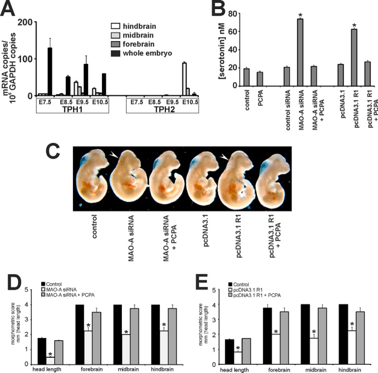
FIGURE 2.
Inhibition of embryonic 5-HT biosynthesis rescues developmental retardations induced by impaired MAO-A expression. A, mouse embryos were dissected (whole embryos and embryonic brain) at different embryonic stages (E7.5–E10.5). Embryonic brains from E8.5 onward were further dissected into fore-, mid- and hindbrain. RNA was extracted, and mRNA levels of TPH isoforms (TPH1 and TPH2) were quantified by qRT-PCR. B–E, mouse embryos were explanted at E7.5 and subjected to in vitro culture. Transfection complexes containing 100 nm siRNA (scrambled siRNA or MAO-A siRNA) or 1 μg/μl plasmid DNA (pcDNA3.1 backbone or pcDNA3.1 R1) were injected into the amniotic cavity. 1 μm PCPA or vehicle was added to the growth medium. After 72 of in vitro culture, embryos were collected and analyzed. B, serotonin levels were quantified as outlined under “Experimental Procedures.” Significances were calculated using Student's t test; n = 6. *, p < 0.001. C, representative images of differently treated mouse embryos are shown. Arrows indicate brain regions with defective development. D and E, developmental progress of the embryos following different treatments was quantified by morphometric scoring. Significances were calculated using Student's t test; n = 4. *, p < 0.001. Error bars represent S.D. If no error bars are visible in the diagrams, no developmental variations between embryos of one experiment could be detected.

