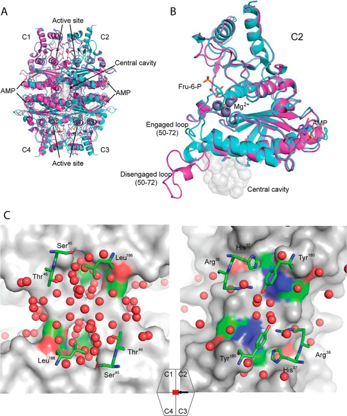
FIGURE 1.
Overview of FBPases. A, alignment of R-state (cyan, product complex, PDB code 1CNQ) and T-state (magenta, AMP complex, PDB code 1EYJ) pFBPases map the locations of AMP binding, active sites, and the central cavity. B, an expanded view of A, showing subunit C2 with loop 50–72 in engaged (cyan) and disengaged (magenta) conformations. C, the central cavity of Leu54 pFBPase (left, IR-state, AMP complex, 1.9 Å, PDB code 1YYZ) and the central region of eFBPase (right, ammonium sulfate complex, 1.5 Å, PDB code 2GQ1). Comparable regions exhibit significant differences in hydration levels. The surface renderings are of subunits C1 and C4 of the tetramer, with ball-and-stick models representing selected residues from subunits C2 and C3 and water molecules (red spheres). The icon with associated arrow indicates the region viewed and viewing direction, respectively. Image generated with PyMOL (57).

