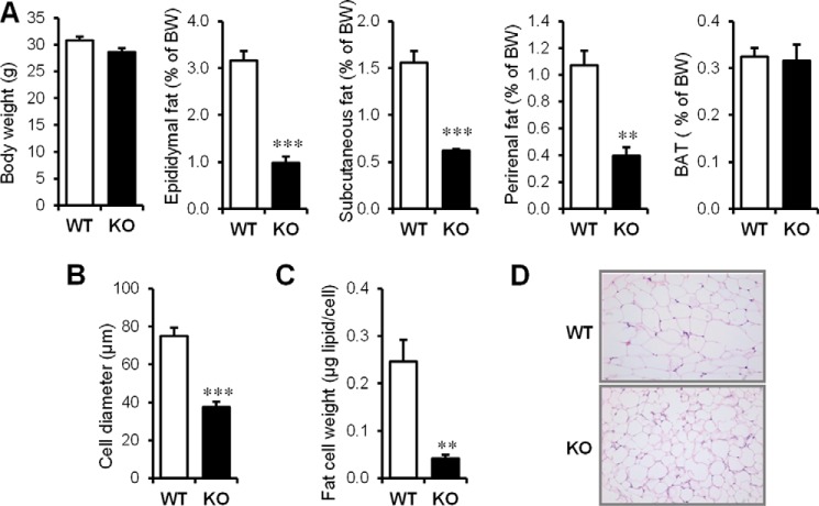
FIGURE 1.
Cavin-1 gene knock-out results in reduced fat pad weight and adipocyte size. A, shown (left to right) is the total body weight and the percent body weight of epididymal, subcutaneous, and perirenal fat pads as well as BAT in 3-month-old male Cavin-1-null mice and their littermates (WT) (n = 5–6). B, average size of isolated adipocytes from 3-month-old Cavin-1-null mice and WT mice. The sizes of 200 randomly selected individual adipocytes from each mouse were determined from scaled images using SPOT software. A total of 1000 adipocytes from 5 mice per genotype were quantified and used for determining the average cell diameter size. C, fat content/cell from the same adipocytes of WT and Cavin-1-null mice is shown. Data are expressed as the mean ± S.E. ***, p < 0.001 for WT versus KO and **, p < 0.01. D, representative H&E staining of epididymal adipose tissue sections from Cavin-1-null (KO) and WT mice is shown at 20× magnification.

