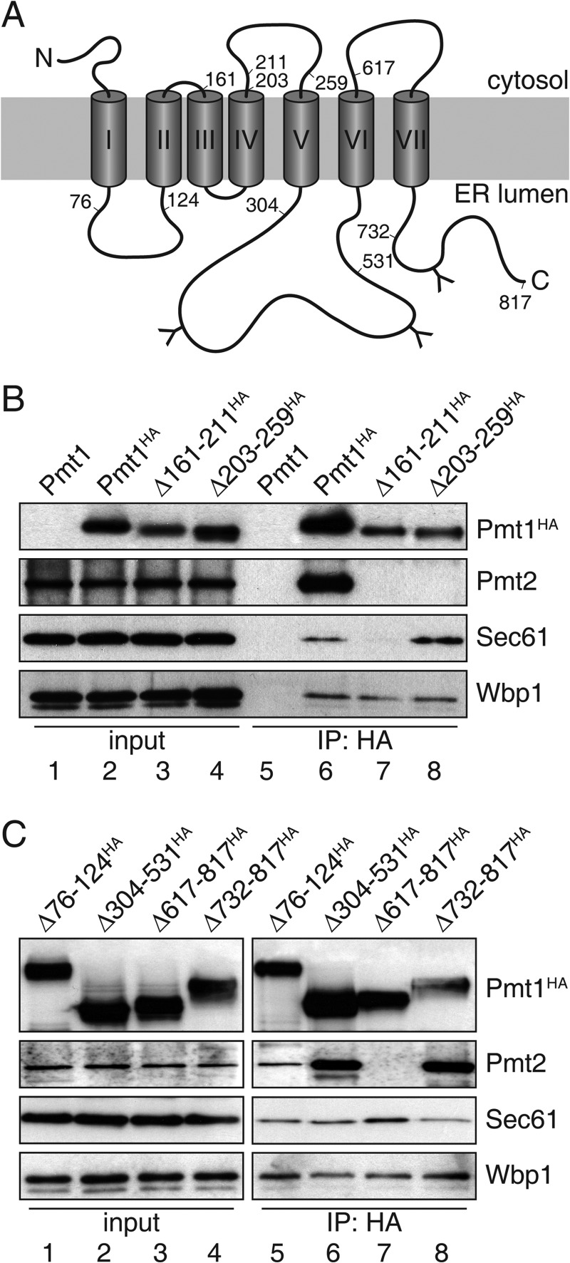
FIGURE 2.
Pmt1 region TMDIII-IV is crucial for association with translocon. A, schematic presentation of Pmt1. Numbers (amino acids positions) indicate deleted regions within Pmt1. Transmembrane spans are depicted as gray cylinders (I–VII) and N-glycosylation sites are indicated by the letter Y. B and C, anti-HA immunoprecipitations were carried out on digitonin-solubilized total membranes from strain pmt1 expressing plasmid-encoded versions of Pmt1 (pSB53, pSB56, pVG9, pVG11, pSB101, pVG13, pSB64, and pSB63; see Table 2). Input lanes of interacting proteins represent 10% of the material used for the IP. Samples were resolved on SDS-polyacrylamide gels and analyzed by Western blotting. Blots were probed with antisera directed against HA-epitope (Pmt1HA), Pmt2, Sec61, or Wbp1 (see Table 3).

