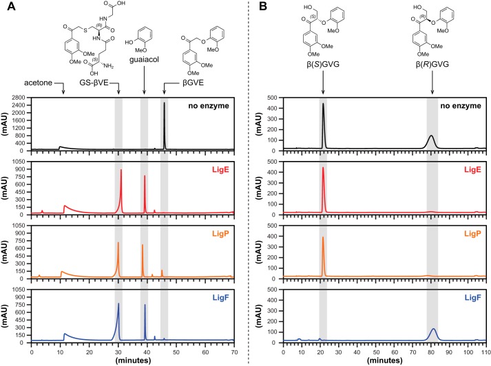
FIGURE 5.
HPLC chromatogram traces of pre-enzyme addition (black), LigE (red), LigP (orange), and LigF (blue) enzyme assay samples. A, C18-reversed phase chromatography of βGVE prior to the addition of and after a 1-h incubation with GSH and either LigE, LigP, or LigF. B, chiral chromatography of racem-βGVG prior to the addition of and after a 1-h incubation with GSH and either LigE, LigP, or LigF.

