Figure 2.
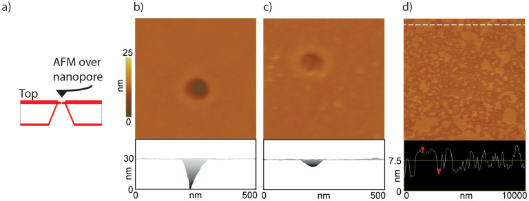
Panel a: Schematic of the AFM-nanopore support imaging setup (not drawn to scale). Panels b and c: Top view and lateral view AFM images of: nanopore alone (without membrane) and the same nanopore covered with DPhPC lipid bilayer reconstituted with Cx43 hemichannels, respectively. The membrane is caved into the pore (possibly due to membrane fluidity and/or by the AFM imaging force. However, the depth of the membrane is only 5–7 nm (lateral view, bottom half of panel c) compared to the uncovered pore (lateral view, bottom half of panel b) where the depth is ~30 nm and is underestimated due to the tip shape and height. Significantly, the membrane, even though bent is stable and intact over several AFM scans. d) AFM image of the lipid bilayer patches (without any reconstituted hemichannels) on the support chip (without the nanopore) The height profile of the patches shown in the bottom half of panel d show the typical bilayer height (5–6 nm) measured by AFM. The center of the image has fewer patches, perhaps representing unfused smaller bilayer patches and/or due to the disruptive action of the AFM tip during multiple scanning in that region. A DPPC lipid was used in this experiment. All images were acquired in contact mode.

