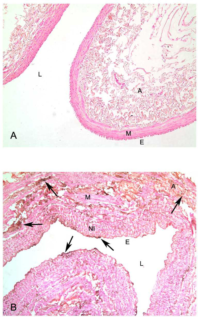
Figure 1.
Panel A. Representative histological section of normal vein. Note thin media (M) and no neointima present. There is also no calcification present after Von Kossa stain. Panel B. Representative histological section of vein obtained at the time of fistula creation with Von Kossa stain. Note presence of calcification (brown and black stain) in endothelium (E), neointima (NI), media (M), and adventitia (A).

