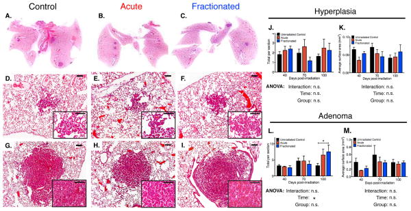
Figure 2. Tumor Burden and Grade in Irradiated K-rasLA1 Lungs Comparable to Unirradiated Controls Seventy Days Post-irradiation.
(A – C) Representative scans of hematoxylin and eosin stained K-rasLA1 lungs seventy days post-irradiation with an acute or fractionated dose of 1.0 Gy 56Fe- particles and age-matched unirradiated controls demonstrate low tumor burden.
(D – I) Representative images of seventy days lungs indicates lesions comprised of only benign lung hyperplasias (D – F) or adenomas (G – I). Inset higher magnification of lesion.
(J – M) Quantification of overall number and size of hyperplasias (J and K) and adenomas (L and M) in K-rasLA1 lungs from forty to one hundred days post-irradiation with an acute or fractionated dose of 1.0 Gy 56Fe- particles and age-matched unirradiated controls (N = 6 – 8 mice per group per timepoint); 3 sections per mouse quantified).
Scale bars denote 100 μm; Graphs show mean ± SEM; Tumor differences determined by two-way ANOVA with Tukey correction; *p < 0.05.

