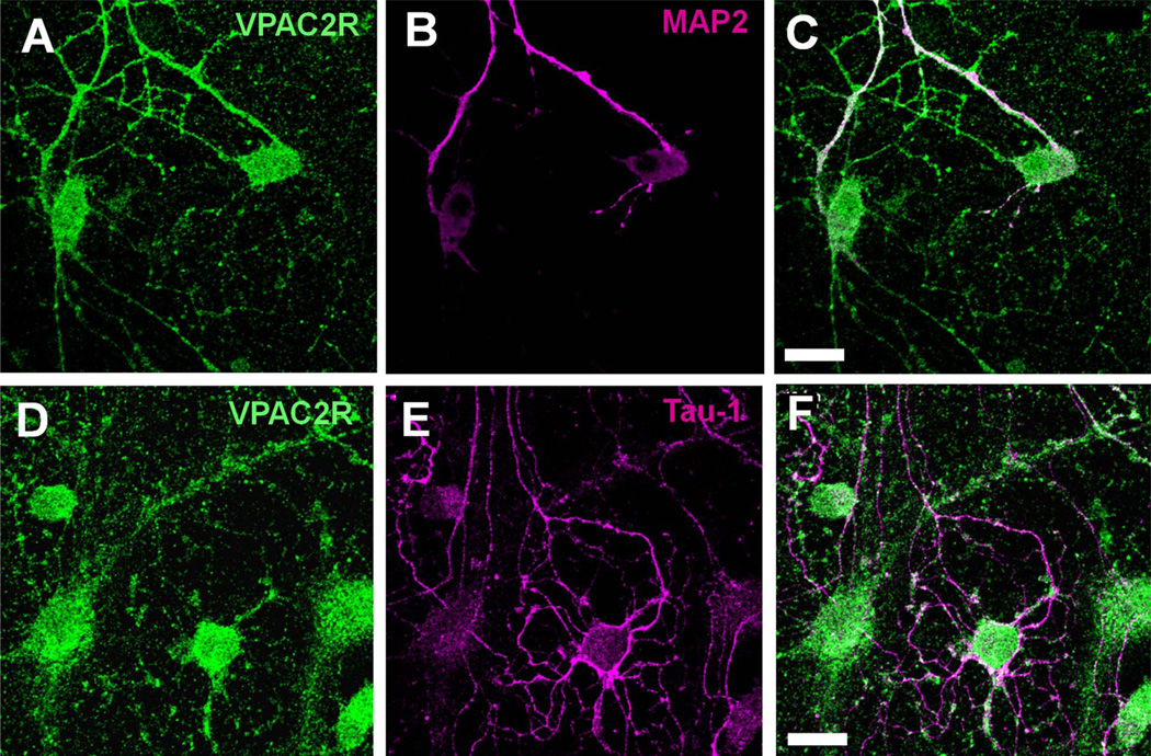
Figure 4.
VPAC2R co-localized with dendritic markers but not with axonal markers in the SCN. A–F: Representative images of dissociated SCN neurons stained for VPAC2R (A,D) and MAP2, a dendritic marker protein (B), or Tau-1, an axonal marker protein (E). Note that VPAC2R (green) staining colocalized with MAP2 (red) along processes (C) but not with Tau-1 (red) staining (F). Scale bar = 20 µm in C (applies to A–C) and F (applies to D–F).

