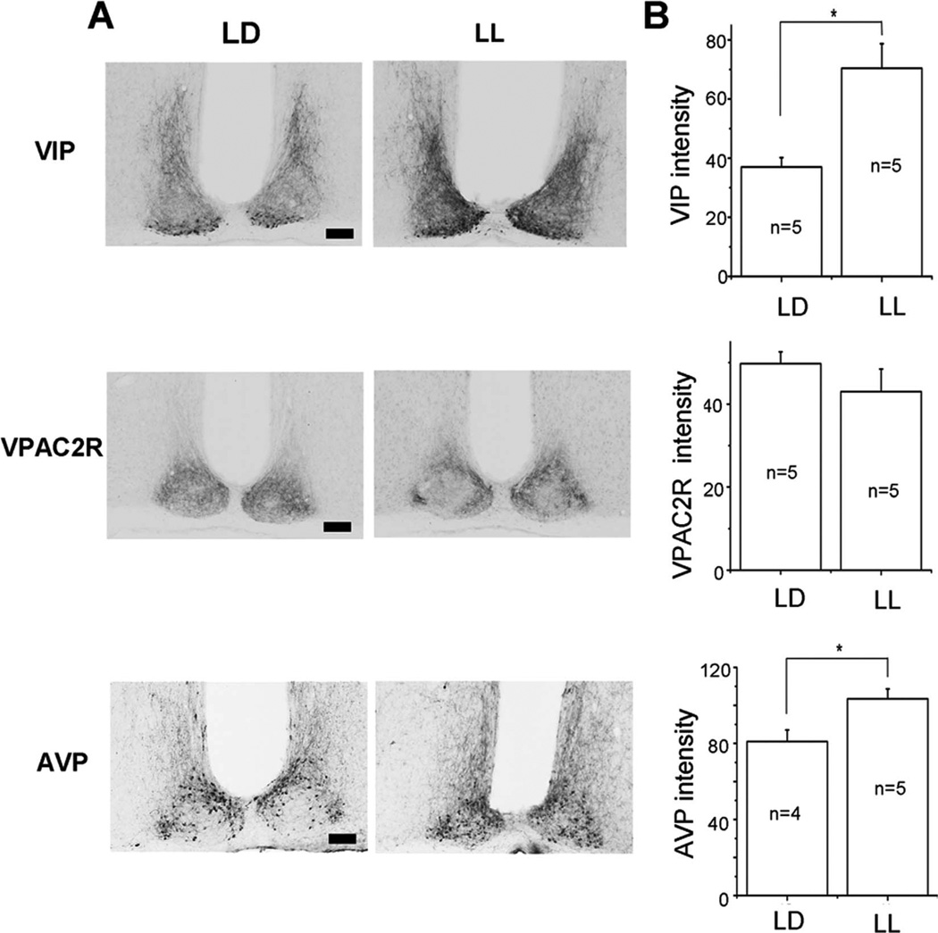
Figure 6.
Constant light increased VIP and AVP, but not VPAC2R expression in the SCN. A: Representative micrographs illustrate VIP-immunoreactive cell bodies in the ventral SCN and their dense projections into the dorsomedial SCN in light–dark (LD; left) and constant light (LL; right). Note the darker projections and cell bodies in LL. Similarly, AVP immunoreactivity increased, whereas VPAC2R labeling did not change in LL B: Mean ± SEM of the staining intensity in the SCN above background for VIP, VPAC2R, and AVP from mice maintained in LD or LL. Asterisks indicate significance, at P < 0.05, and n indicates the number of mice used. Scale bar = 100 µm in A.

