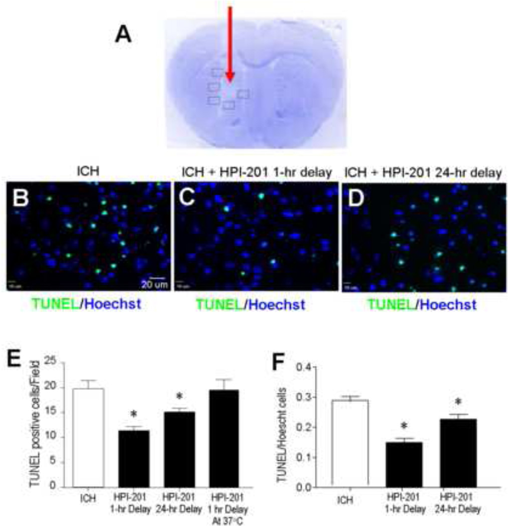
Figure 2. Intracerebral hemorrhage-induced brain damage, cell death and effect of pharmacological hypothermia.
A. Nissl staining revealed tissue damage in the hematoma area 3 days after autologous blood injection (arrow). Boxed frames show the location of assay areas of TUNEL-positive cells. No similar tissue damage was seen in contralateral hemispheres. B - D. TUNEL staining of DNA damage and cell death in the perihemorrhage region of brain sections 48 hrs after ICH. Immunofluorescent images showing TUNEL-positive cells (green) and total cells stained with Hoechst 33342 (blue) in the three experimental groups: ICH control, ICH plus 1 hr delayed treatment with HPI-201 and ICH plus 24 hr delayed treatment with HPI-201. E. The number of TUNEL-positive cells in randomly selected survey fields. HPI-201 treatment initiated either 1 hr or 24 hr after ICH significantly decreased the amount of cell death. As a control that was performed after the initial experiments, additional animals that received HPI-201 injection were kept at 37°C using a heating blanket with feedback control (see Figure 1). These animals showed similar cell death as ICH only controls. F. The ratio of TUNEL-positive cells against total cells was compared in hemorrhage mice that received saline or delayed HPI-201 treatments. *. p<0.05 vs. ICH control; n=7 and 8 in 1 hr and 24 hr delay groups, respectively; n=7 in HPI-201 at 37°C group. All ICH control animals received saline injection in this investigation.

