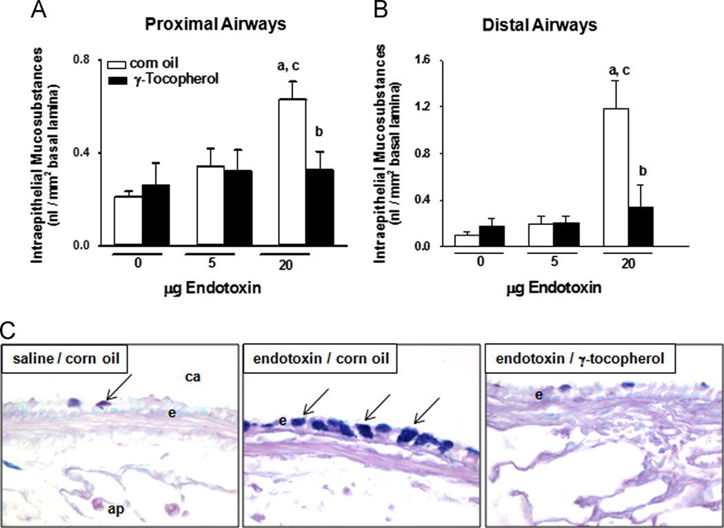
Fig. 3.
Mucous cell metaplasia. Intraepithelial mucosubstances in (A) proximal and (B) distal conducting airways were quantified in lung sections by morphometric approaches as described under Materials and methods. a, significantly different from respective group given 0 µg endotoxin; b, significantly different from respective group given corn oil; c, significantly different from respective group given 5 µg endotoxin; p < 0.05. (C) Mucosubstances were histochemically detected in AB/PAS-stained tissues as blue positive intracellular staining (arrows) of airway epithelium. ca, conducting airway; e, airway epithelium; ap, alveolar parenchyma. (For interpretation of the references to color in this figure legend, the reader is referred to the web version of this article).

