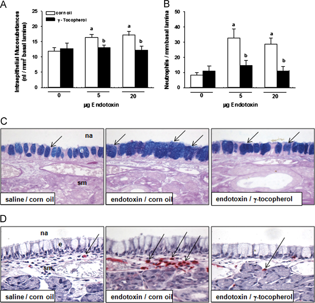
Fig. 6.
Nasal responses. Mucous cell metaplasia was determined morphometrically as increases in (A) intraepithelial mucosubstances in (C) the respiratory epithelium lining the nasal septum stained with AB/PAS, which appear as intracellular blue-stained material (arrows). (B) Submucosal neutrophils were enumerated by morphometric approaches in nasal septum in immunohistochemically treated tissue to detect (D) cells staining positive (arrows, red cells) for anti-rat neutrophil antibody, as described under Materials and methods. a, significantly different from respective group given 0 µg endotoxin; b, significantly different from respective group given corn oil; p < 0.05. na, nasal airway; e, epithelium, sm, submucosa. (For interpretation of the references to color in this figure legend, the reader is referred to the web version of this article).

