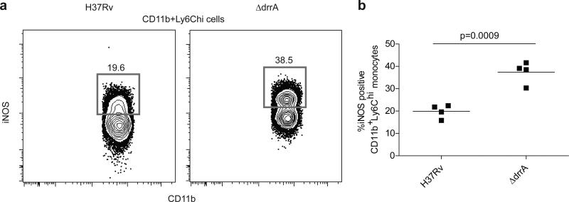
Figure 3. Elevated frequencies of iNOS expressing inflammatory monocytes in mice infected with PDIM-deficient M. tuberculosis.
C57B6 mice were infected via the aerosol route with H37Rv or an isogenic PDIM-deficient mutant (ΔdrrA). Lung tissue was harvested on 21 DPI and iNOS (protein expression was measured via flow cytometry. Representative FACS plots (a) and graphical depiction (b) of frequencies of iNOS expressing cells within the Ly6C+CD11b+ inflammatory monocyte population. Representative of two separate experiments. Student's unpaired t test.

