Figure 3.

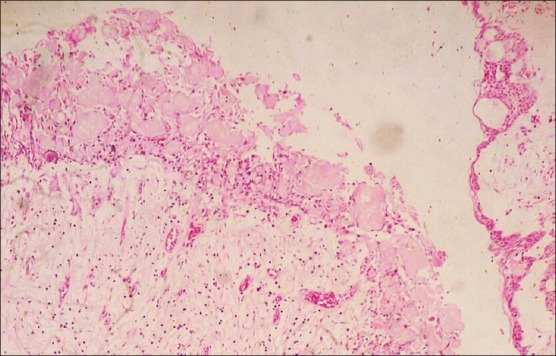
Photomicrograph showing cyst epithelium with numerous ghost cells and few calcified masses. Basal cells are cuboidal to columnar in shape showing hyperchromatism, few showing reverse polarity with palisading appearance (H and E, ×10)

Photomicrograph showing cyst epithelium with numerous ghost cells and few calcified masses. Basal cells are cuboidal to columnar in shape showing hyperchromatism, few showing reverse polarity with palisading appearance (H and E, ×10)