
Fig. 5.
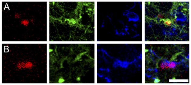
Phenotype of BrdU-labeled cells in the amygdala. Confocal laser microscope images display labeling for BrdU (red; left), TuJ1 (green; center left), NG2 (blue; center right), and all three markers (right) in the posterior cortical nucleus (pCorA) of the amygdala in the vole brain. Some BrdU-labeled cells coexpressed the neuronal (TuJ1; A) or glial progenitor (NG2; B) marker. Scale bar – 5 μm in B (applies to A–B, all panels).