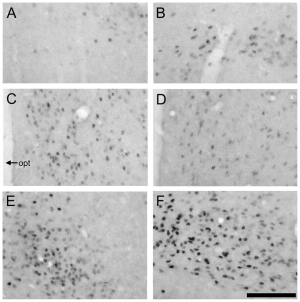
Fig. 6.
Photomicrographs displaying ERα labeling in the amyg-dala and ventromedial hypothalamus (VMH) in prairie (left) and meadow (right) voles. In the posterior cortical nucleus of the amyg-dala (A,B) and VMH (E, F), meadow voles (B, F) appeared to have more ERα-labeled cells than did the prairie voles (A, E). However, no species differences were found in the posterior medial nucleus of the amyg-dala (C, D). opt, optic tract. Scale bar – 200 μm in F (applies to A–F).

