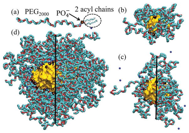
FIG. 1.
(a) DSPE-PEG2000 monomer with Na+ counterion. (b) Equilibrated SSM-10 in water. The characteristic layers in this system are alkane core (rcore ≈ 0 – 1.2 nm, shown as a gold surface), ionic interface (rint ≈ 1.2 – 1.8 nm; the negatively charged groups are shown as black and orange clusters), PEG corona (rPEG ≈ 1.8–3.0 nm, shown as turquoise and red chains), and aqueous ionic solution (raq > 3.0 nm, not shown for clarity). (c) Equilibrated SSM-20 in water, modeled after the SSM observed by DLS experiments in PB buffer (low ionic strength solution), with rcore ≈ 0 – 1.7 nm, rint ≈ 1.7 – 2.3 nm, rPEG ≈ 2.3 – 3.8 nm, and raq > 3.8 nm. (d) Equilibrated SSM-90 in 0.16 M NaCl solution, with rcore ≈ 0 – 2.4 nm (along minor axis), rint ≈ 2.4 – 3.0 nm, rPEG ≈ 3.0 – 7.5 nm, and raq > 7.5 nm. In SSM shown in (c–d), groups and PEG chains are removed for clarity on the left sides of the images.

