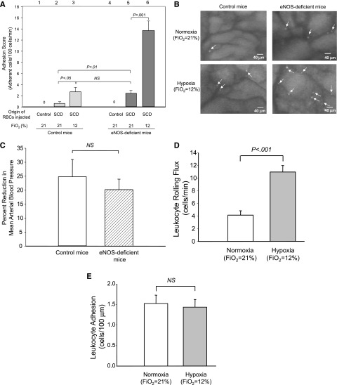
Figure 3.
Adhesion of sRBC and leukocytes in control and eNOS-deficient mice under normoxia (procedure I) or hypoxia (procedure II). (A) Analysis of RBC adhesion to endothelial cells by intravital microscopy. Labeled RBCs were prepared from either control or SCD mice and injected into control or eNOS-deficient mice. Note that RBCs prepared from control mice did not adhere to endothelial cells in either control or SCD mice, suggesting that the intravital microscopy analysis eliminates false-positive cell–cell interactions. Lanes are shown at the top of figure, and the origins of injected RBCs and oxygen tensions are shown at the bottom of figure. Values were mean ± SEM obtained from 3 to 7 mice in each group. P values are shown at the top of the figure. (B) Frame-captured images from videotaped intravital microscopy of bone marrow venules in control (left panels) and eNOS-deficient mice (right panels) after injection of BCECF-labeled sRBCs under normoxia (upper panels) and hypoxia (low panels). Arrows indicate adhered sRBCs. (C) Percent reduction in MABP in response to hypoxia (Fio2 = 12%) in control and eNOS-deficient mice. Values were mean ± SEM from 5 to 7 mice in each group. (D-E) Leukocyte rolling (D) and leukocyte adhesion (E) to endothelial cells in eNOS-deficient mice at normoxia (procedure I) or at hypoxia (procedure II). PE rat anti-CD45 antibody was injected into eNOS-deficient mice. Leukocyte adhesion was quantified by counting the number of adherent cells (stationary for >30 seconds) in a 100-μM length of the vessel. Note that only leukocyte rolling is increased at the onset of hypoxia. Values were mean ± SEM from 5 to 7 mice in each group. P values are shown in the figure.

