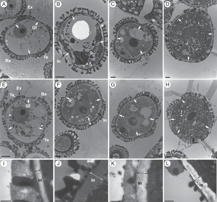
Fig. 7.
Transmission electron micrographs of pollen development in the bcmf8 lines. (A–D) Micrographs of pollen from empty vector pBI121-transformed plants (CK) showing normal pollen morphology with three apertures and a normal development process from the early uninucleate microscope stage to the mature pollen stage. The pollen at the early uninucleate (A), late uninucleate (B), binucleate (C) and trinucleate (D) microscope stages was observed. (E) Micrograph of pollen from the bcmf8 lines showed normal morphology with three apertures at the early uninucleate microscope stage. (F) Micrograph of pollen from the bcmf8 lines at the late uninucleate microscope stage showing a remarkable thickening in intine (black arrow) both inside and outside the aperture region. (G) Micrograph of pollen from the bcmf8 lines at the binucleate microscope stage showing the number of potential apertures to increase to four instead of three as normal. (H) The uneven distribution of the apertures in the bcmf8 pollen at the mature pollen stage. (I) Mature intine outside the aperture region of the control pollen. (J) Mature intine outside the aperture region of the bcmf8 pollen. There was a remarkable thickening outside the aperture region, compared with the control pollen. (K) Mature intine inside the aperture region of the control pollen. (L) Mature intine inside the aperture region of the bcmf8 pollen. Gf, aperture; In, intine; Ex, exine; Te, tectum; Ba, baculum. Scale bars = 2 µm (A–H); 0·5 µm (I–L).

