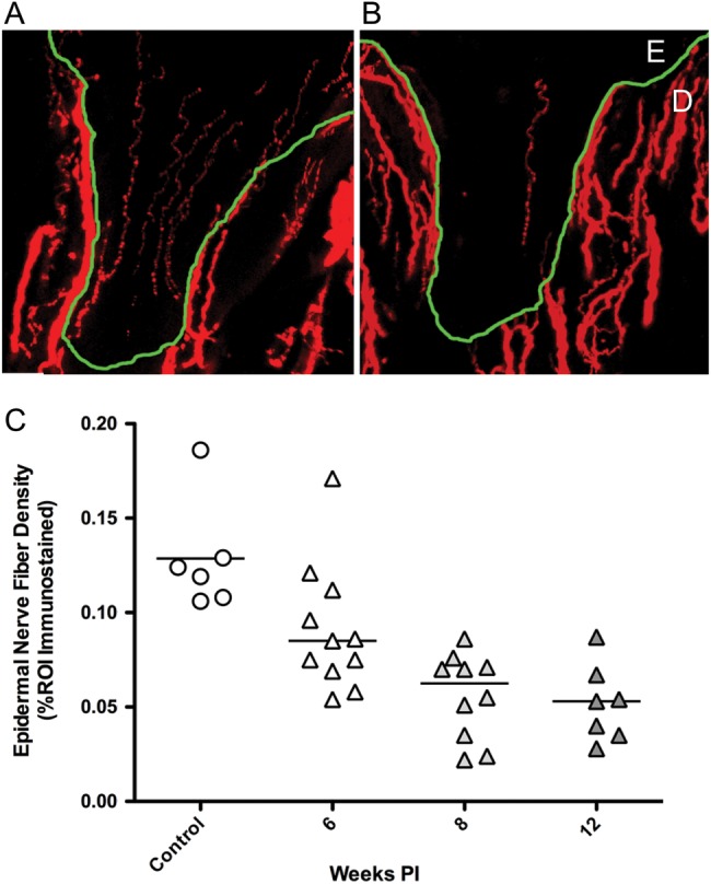
Figure 4.

Epidermal nerve fiber density is reduced with SIV-infection. PGP9.5 immunostained sections of footpad skin from an uninfected control (A) and SIV-infected macaques (B) showed marked decline in ENF density. Nerve fibers (red) are present in the epidermis (E) and dermis (D), with the dermal-epidermal junction traced in green. (C) Measurement of ENF density demonstrated reduced ENF density in SIV-infected macaques (triangles) compared to uninfected controls (circles), with significant decrease developing by eight weeks p.i., then declining further at the twelve week time point. (P < 0.001, analysis of variance). Reprinted with permission from Laast et al. 2011).
