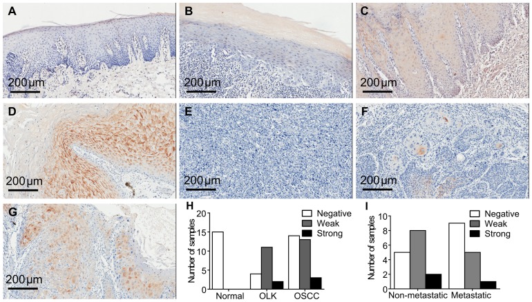
Figure 1. Immunostaining of hBD-1 protein in different tissue samples.
A) Examples of typical images of hBD-1 in normal oral mucosa (negative); B) OLK (negative); C) OLK (weak); D) OLK (strong); E) OSCC (negative); F) (weak); G) OSCC (strong). H) Number of samples with negative, weak and strong staining of hBD-1 in normal oral mucosa, OLK and OSCC. I) Percentage of negative, weak and strong staining of hBD-1 in OSCC with or without metastasis.

