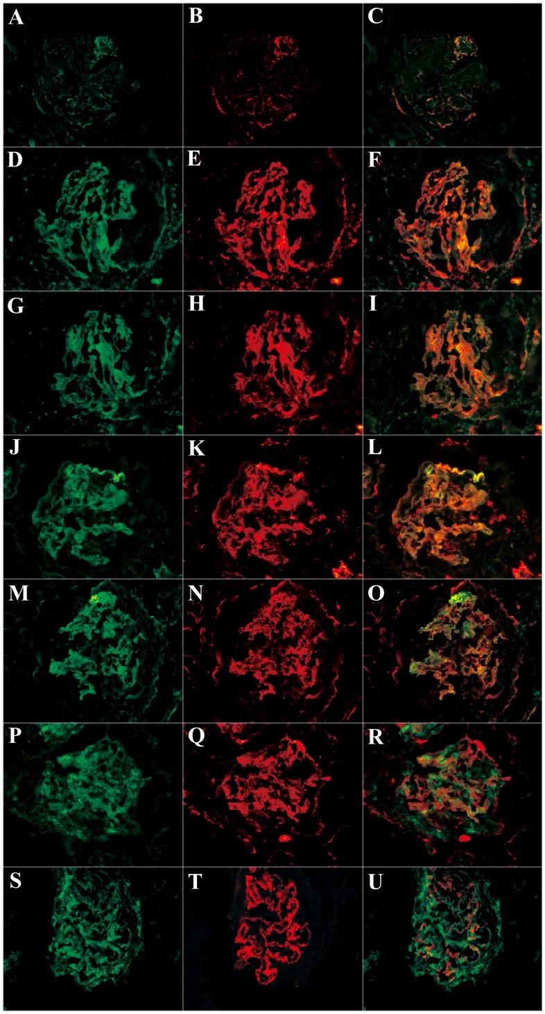
Figure 3. Co-localization of various complement components were detected in frozen sections by immunofluorescence using laser confocal microscopy (magnification ×400).
A: IgG linear deposition along the glomerular capillary wall; B: C3d granular deposition along the glomerular capillary wall in the same section; C: IgG and C3d co-localized completely; D: C1q linear deposition along the glomerular capillary wall; E: C5b-9 granular deposition along the glomerular capillary wall in the same section; F: C1q and C5b-9 co-localized completely; G: factor B linear deposition along the glomerular capillary wall; H: C5b-9 granular deposition along the glomerular capillary wall in the same section; I: factor B and C5b-9 co-localized completely; J: properdin linear deposition along the glomerular capillary wall; K: C5b-9 granular deposition along the glomerular capillary wall in the same section; L: properdin and C5b-9 co-localized completely; M: properdin linear deposition along the glomerular capillary wall; N: C3d granular deposition along the glomerular capillary wall in the same section; O: properdin and C3d co-localized completely; P: MBL diffusive deposition; Q: C5b-9 granular deposition along the glomerular capillary wall in the same section; R: MBL and C5b-9 could not co-localize; S: MBL diffusive deposition; T: C4d granular deposition along the glomerular capillary wall in the same section; U: MBL and C4d partially co-localized along the glomerular capillary wall.

