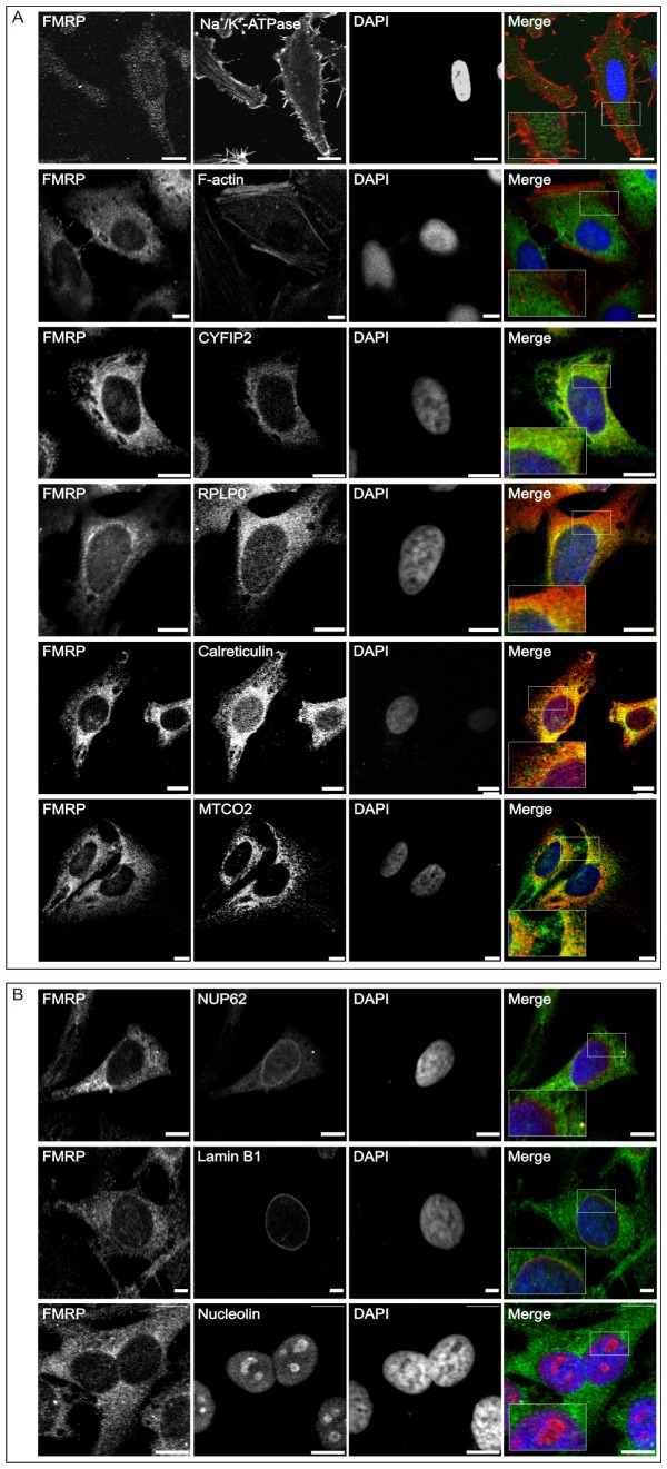
Figure 1. FMRP is localized at various intracellular sites in HeLa cells.
Confocal laser scanning microscopy (cLSM) images of HeLa cells depicting endogenous FMRP (green channel) costained with various cytosolic (A) and nuclear (B) markers (red channel), including antibodies against CYFIP2, RPLP0 (ribosomal proteins), nucleolin (nucleolar marker), MTCO2 (mitochondrial protein), NUP62 (nucleoporins), lamin B1 (nuclear intermediate filament proteins), and calreticulin (endoplasmic reticulum marker). Detection of Na+/K+-ATPase and phalloidin staining were used to detect the cellular membrane and F-actin, respectively. DNA was stained by using DAPI (blue channel). Boxed areas in the merged panels depict enlarged areas of interest. Scale bar: 10 μm.

