
Figure 2. Illustrated T1W MRI post-contrast enhancement of trismus patient after RT.
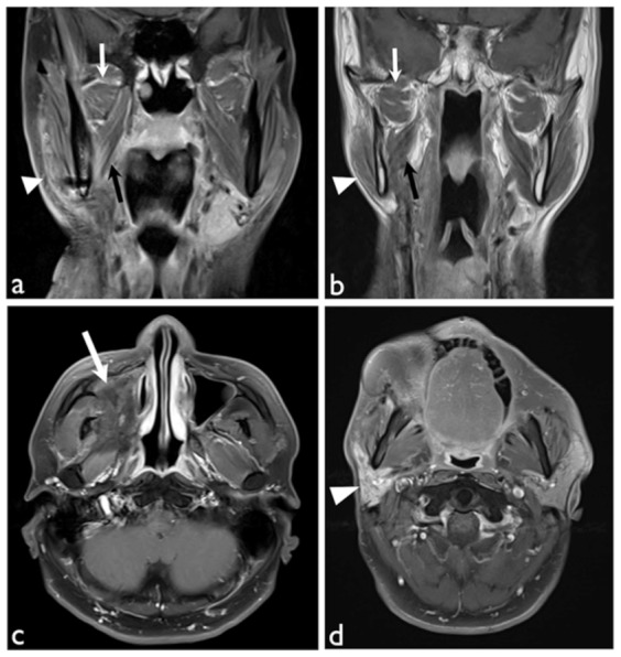
(a) Coronal view with fat saturation showed radiation effects in right lateral (white arrows), medial (black arrows) pterygoid muscles and masseter muscle (white arrowhead) with increased enhancement. (b) Coronal view showed atrophic change of the right lateral (white arrows), medial (black arrows) pterygoid muscles and masseter muscle (white arrowhead). (c) Axial view with fat saturation showed remarkable fibrotic tissue (white arrow) occupying right maxillary sinus and pterygoid space. (d) Axial view with fat saturation showed increased enhancement with atrophic change of the right parotid gland. All these findings were rated as point 2 according to our Likert scoring system of signal abnormality (SA score).