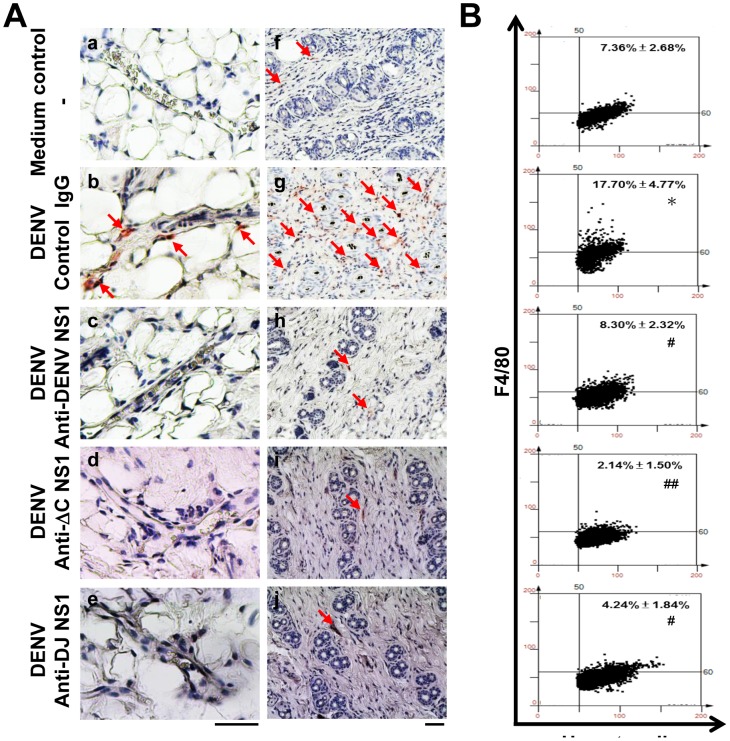
Figure 7. Anti-DENV NS1, anti-ΔC NS1 and anti-DJ NS1 Abs reduce macrophage infiltration in DENV-infected mice.
(A) Hematoxylin-based nuclear staining followed by immunohistochemical staining of F4/80, a macrophage marker, in skin vessel (a–e) and dermis layer (f–j) of treated mice were observed. The arrows indicate positive staining. (Magnification: ×200; bar = 100 μm) (B) Quantification of F4/80 staining was performed on skin sections using HistoQuest analysis software. A representative scattergram plot of each group is shown and the percentage of F4/80 positive cells is shown as means ± SD obtained from three mice of each group. * P<0.05 as compared with medium control group. # P<0.05; ## P<0.01 as compared with DENV plus control IgG group.

