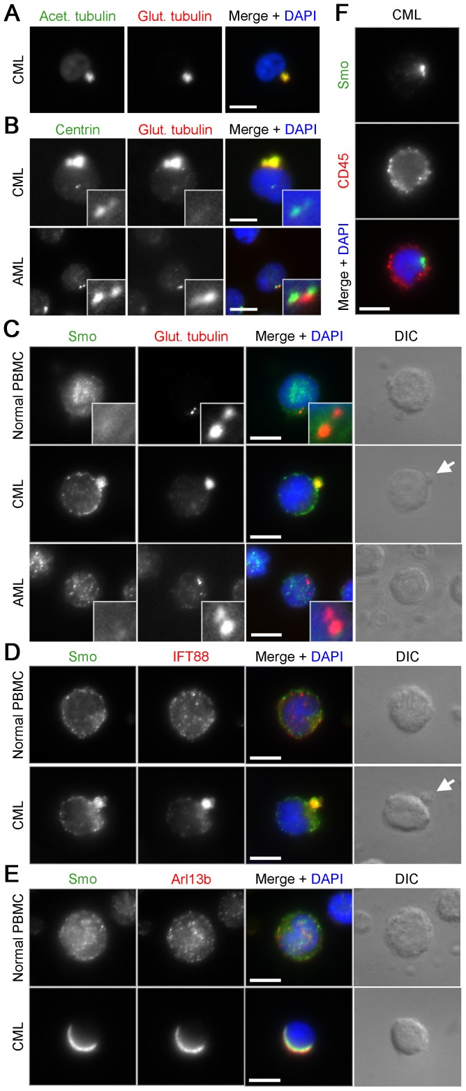
Figure 5. Localization of ciliary proteins in human CML cells.
(A) Human CML cells stained with antibodies against acetylated (green) and glutamylated tubulin (red). (B) CML and AML cells stained with antibodies against centrin (green) and glutamylated tubulin (red). (C) Normal PBMC, CML, and AML cells stained with antibodies against Smoothened (Smo) (green) and glutamylated tubulin (red). (D) CML cells stained with antibodies against Smo (green) and either IFT88 (red). (E) CML cells stained with antibodies against Smo (green) and Arl13b (red). DNA is stained using DAPI (blue), scale bars: 5 μm, insets: 10× magnification, white arrows: cell protrusions. In each case >100 cells were imaged, and the phenotype represented in the images shown was seen in greater than 50% of cells.

