Figure 3.

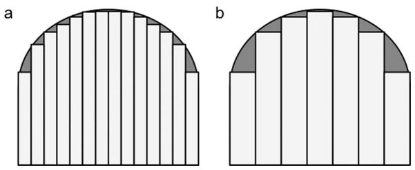
Schematic images of the femoral condyle cartilage segmentation on (a) thin and (b) thick slice images. The area of underestimation on thick slice images (gray area) is larger than that on thin slice images.

Schematic images of the femoral condyle cartilage segmentation on (a) thin and (b) thick slice images. The area of underestimation on thick slice images (gray area) is larger than that on thin slice images.