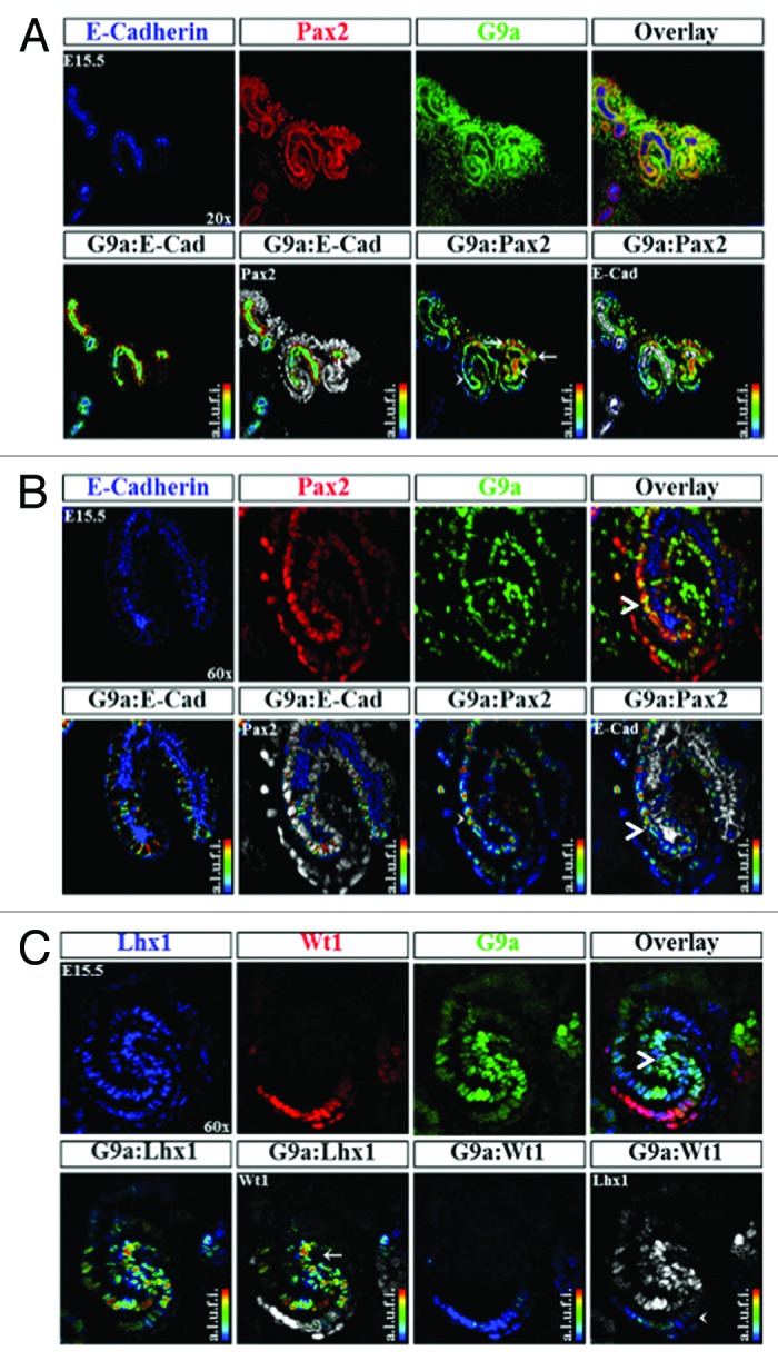
Figure 9. Distribution of H3K9 KMT, G9a, in the developing kidney. (A) G9a is expressed in epithelial (arrowhead) and mesenchymal components (arrows) ×20. (B) A higher power view (×60) showing G9a expression in an S-shaped body and its relative enrichment at the junction of the proximal and distal segments (arrowhead). (C) Co-staining of G9a with Lhx1 (a marker of nascent nephrons) and WT1 (a marker of podocytes) reveals G9a expression at the junction of proximal and distal segments of S-shaped body (arrow and arrowhead).
