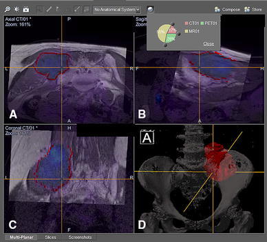
Fig. 2.

CT/MR/PET fusion images are shown in the navigation display in a patient with a left pelvic tumor involving the posterior superior iliac crest and sacral ala. Wide resection was performed under navigational guidance via a posterior approach, and the left L5 and S1 nerve roots were preserved. Different proportions of image modality could be adjusted on the axial (a), reformatted sagittal (b), and coronal (c) views of the fused images. d A 3D bone tumor model was created after the tumor extent was outlined on the MR images. Surgeons then could accurately define the resection planes after studying the fused 2D images and 3D model
