
Fig. 3.
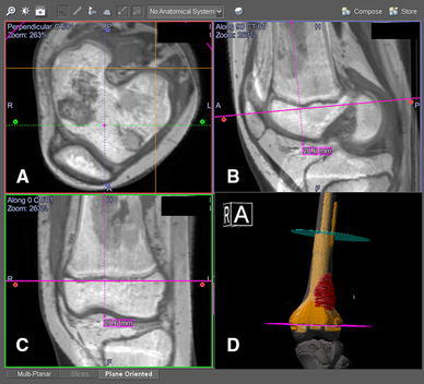
CT/MR fusion images are shown in the navigation display in a 13-year-old girl with a left distal femoral osteosarcoma. Joint-preserving resection and reconstruction were performed with a custom CAD prosthesis. The intraosseous tumor extent was better seen on the T1-weighted MR images. The resection plane (pink) was defined as 2 cm proximal to the knee joint. The resection level could be checked with the axial (a), reformatted sagittal (b), and coronal (c) views of the fused images. CAD data of the joint-preserving prosthesis also could be imported into the navigation planning and seen on the 3D model (d) with the resection planes. Surgeons checked the final design of the CAD prosthesis before giving approval to the manufacturer