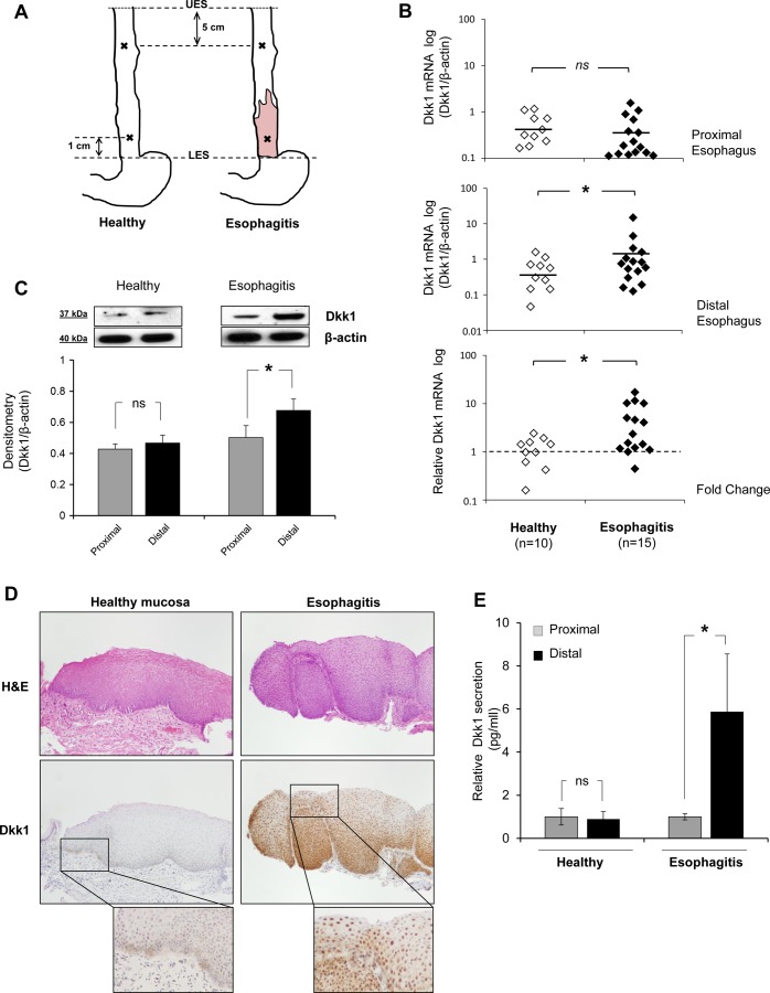
Fig. 1.
Dkk1 expression in healthy esophageal mucosa and reflux esophagitis tissue. A: topography of biopsy acquisition. UES and LES, upper and lower esophageal sphincter. B: scatter graph of quantitative RT-PCR data for Dickkopf-1 (Dkk1) mRNA expression in distal and proximal esophageal mucosa of healthy individuals (n = 10) and esophagitis patients (n = 15). Data were normalized to β-actin expression. Top: Dkk1 mRNA expression in proximal biopsies of the 2 groups is not different. Middle: Dkk1 mRNA expression is greater in biopsies from esophagitis patients than healthy controls. *P ≤ 0.05 (by Mann-Whitney test). Horizontal lines indicate means. Bottom: fold change in expression of Dkk1 between proximal and distal mucosa. Biopsies from esophagitis patients show greater differential expression between proximal and distal mucosa than biopsies from healthy controls. *P ≤ 0.05 (by Mann-Whitney test). C: Western blot analysis of Dkk1 protein expression in human esophageal biopsies. Top: representative blots. Bottom: Dkk1 protein expression relative to β-actin expression in biopsies from healthy controls (n = 3) and esophagitis patients (n = 8). Values are means ± SE. *P ≤ 0.05 (by paired t-test); ns, not significant. D: immunohistochemistry for Dkk1 in esophageal biopsies. Sections were stained with hematoxylin-eosin (H&E) for histological confirmation of esophagitis. Images represent results from 4 experiments. E: ELISA of Dkk1 protein secretion in organ culture medium from distal mucosa relative to paired proximal mucosa in healthy controls (n = 3) and esophagitis patients (n = 3). Values are means ± SE. *P ≤ 0.05 (by paired t-test).

