Fig. 1.
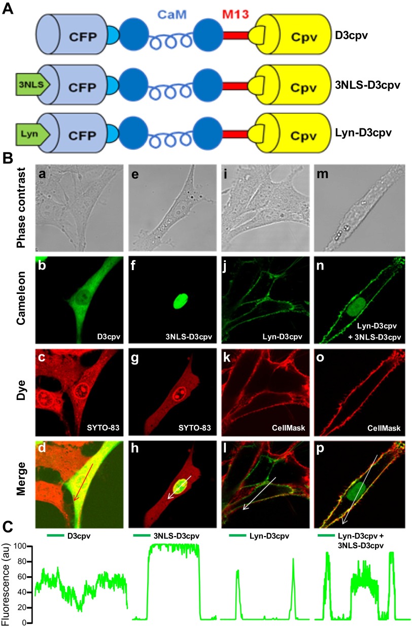
Subcellular distribution of D3cpv, 3NLS-D3cpv, and Lyn-D3cpv in transiently transfected rat pulmonary arterial smooth muscle cells (PASMCs). A: schematic structure of the D3cpv, 3NLS-D3cpv, and Lyn-D3cpv cameleon probes. D3cpv containing improved calmodulin (CaM) and M13 sequences (38) (top) was modified by inserting 3NLS (middle) and Lyn (bottom) sequences at the NH2 terminus. CFP, cyan fluorescent protein. B: confocal images of rat PASMCs transfected with D3cpv (a–d), 3NLS-D3cpv (e–h), Lyn-D3cpv (i–l) and cotransfected with 3NLS-D3cpv and Lyn-D3cpv (m–p). Phase contrast images (a, e, i, and m), cameleon cpV fluorescence (b, f, j, and n), SYTO 83 Orange staining (c and g), and CellMask Orange plasma membrane staining (k and o) are shown. Overlay images of b and c (d), f and g (h), j and k (l), and n and o (p) are indicated. C: linear fluorescence profiles of cpV fluorescence distribution in the cells across the arrow. au, Arbitrary units.

