Fig. 6.
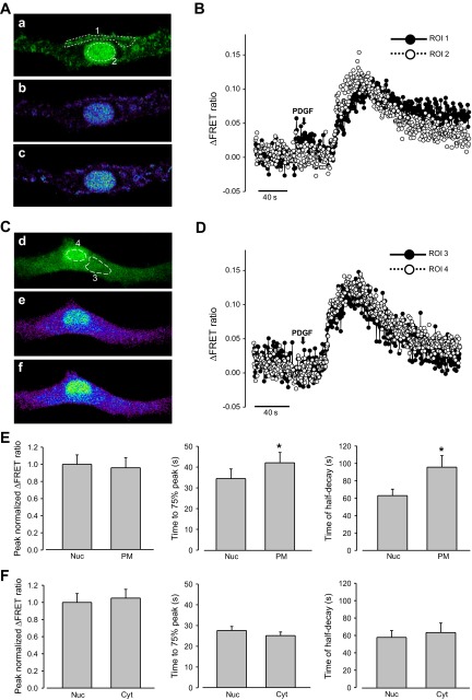
Effects of platelet-derived growth factor (PDGF) on PM, cytoplasmic, and nucleoplasmic Ca2+ increase in rat PASMCs. A: 2D confocal images of a PASMC cotransfected with Lyn-D3cpv and 3NLS-D3cpv, showing cpV fluorescence (a), and Ca2+ mobilization in the PM and nucleoplasm before (b) and after (c) PDGF (20 ng/ml) treatment. B: ΔFRET fluorescence ratio traces showing Ca2+ mobilization in the PM (ROI 1) and nucleoplasm (ROI 2) of rat PASMCs (from A) after PDGF treatment. C: 2D confocal images of a PASMC cotransfected with D3cpv and 3NLS-D3cpv, showing cpV fluorescence (a), and Ca2+ mobilization in the cytoplasm and nucleoplasm before (b) and after (c) PDGF treatment. D: ΔFRET fluorescence ratio traces showing Ca2+ mobilization in the cytoplasm (ROI 3) and nucleoplasm (ROI 4) of rat PASMCs (from C) after PDGF treatment. E: statistical analysis of the peak normalized ΔFRET fluorescence ratio, time to 75% of peak, and time of half-decay of PDGF-induced Ca2+ transients in the nucleoplasm and PM of rat PASMCs. F: statistical analysis of the normalized ΔFRET fluorescence ratio peak intensity, time to 75% of peak, and time of half-decay of PDGF-induced Ca2+ transients in the nucleoplasm and cytoplasm of rat PASMCs. Paired t-test was used to calculate P values. *P < 0.05 compared with nucleoplasmic. Error bars denote SE.

