
Fig. 7.
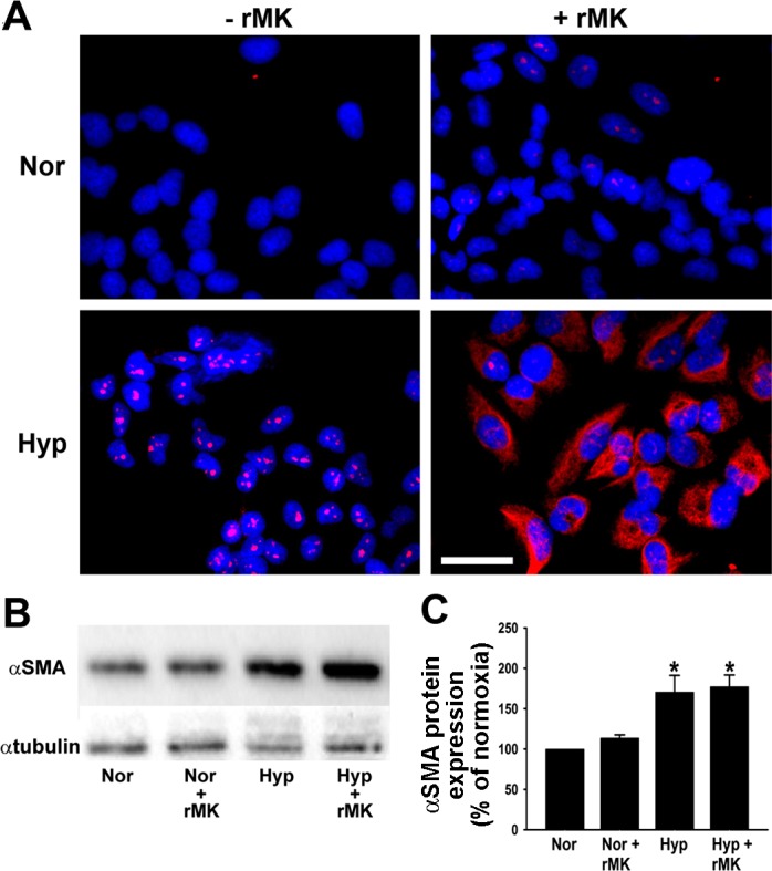
Expression of α-smooth muscle actin (α-SMA), a mesenchymal cell marker, is increased in hypoxic A549 cells by rMK. A: serum-starved A549 cells were treated with rMK (100 ng/ml) and exposed to either normoxia or hypoxia for 72 h. At the end of the exposure, α-SMA (red) was immunodetected. Nuclei are blue. Representative micrographs from 1 of the 3 independent experiments are shown. Scale bar = 10 μm. B: increase in α-SMA protein levels as observed on a representative Western immunoblot. Serum-starved epithelial cells were treated with rMK (100 ng/ml) and then exposed to either normoxia or hypoxia for 72 h before Western immunoblotting for detection of α-SMA and α-tubulin. C: quantification of α-SMA levels from 3 different immunoblots (e.g., Fig. 7B) in response to rMK treatment of normoxic and hypoxic cells. *P < 0.01, compared with Nor and Nor + rMK results.