
Fig. 8.
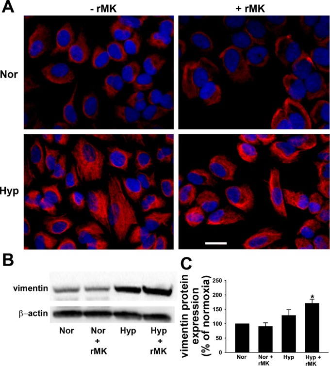
rMK induces vimentin expression in hypoxic A549 cells. A: immunofluorescent staining for vimentin (red). A549 cells were grown with or without rMK and exposed to either normoxia or hypoxia for 72 h. Representative photomicrographs from 3 independent experiments are shown. Scale bar = 10 μm. B: Western immunoblot showing vimentin expression. Serum-starved A549 cells were exposed to either normoxia or hypoxia in the absence or presence of rMK (100 ng/ml). After 72 h of exposure, Western immunoblots for detection of vimentin and β-actin were performed. A representative immunoblot from 3 independent experiments is shown. C: quantification of vimentin levels from 3 Western immunoblots (e.g., Fig. 8B) in normoxic and hypoxic cells in the presence or absence of rMK. *P < 0.01, compared with Nor and Nor + rMK.