
Fig. 4.
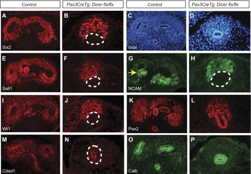
Immunofluorescent staining of E12.5 kidneys shows a failure in ureteric branching and fewer nephron progenitors in Pax3CreTg, Dicerflx/flx kidneys. A, B, E–N: although Six2, Sall1, NCAM, Wt1, Pax2, and Cited1 continues to be present in Pax3CreTg, Dicerflx/flx nephron progenitors, there are far fewer positive cells than in controls (A, E, G, I, K, M, O). C and D: labeling of nuclei with DAPI confirms the histological presence of nephron progenitors and the ureteric bud. O and P: calbindin staining reveals unbranched ureteric buds in Pax3CreTg, Dicerflx/flx kidneys compared with branches seen in controls. Yellow arrow, renal vesicle; white dashed lines, ureteric bud. The magnification is ×20.