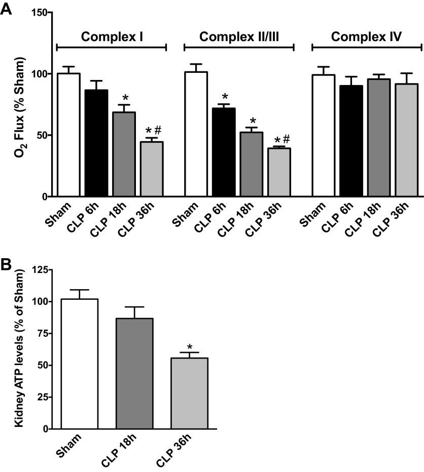
Fig. 1.
Sepsis caused a decline in renal mitochondrial complex respiration and ATP production. A: high-resolution respirometry (HRR) was used to assess the status of mitochondrial complexes [I, II/III, and IV of the electron transport chain (ETC)] of fresh renal biopsies harvested 6–36 h post-cecal ligation and puncture (CLP). Values are oxygen flux expressed as a percentage of sham levels (means ± SE, n = 4–8 mice/group). B: ATP levels in the kidney at 18 and 36 h are presented as percentage of sham levels (means ± SE, n = 7–8 mice/group). *P < 0.05 compared with Sham and #P < 0.05 compared with CLP 18 h.

