
Fig. 2.
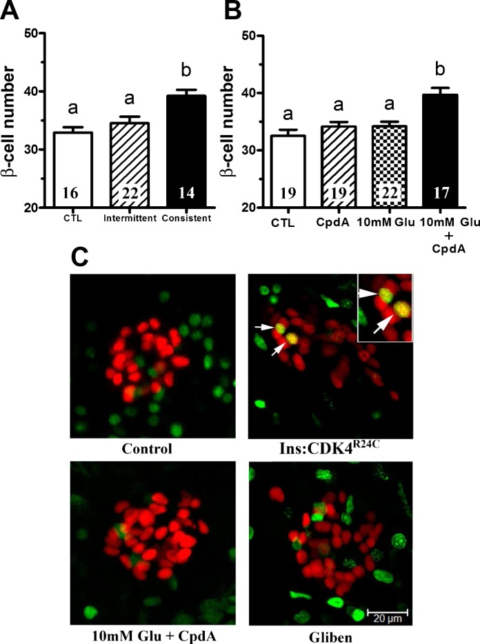
Pharmacological activation of KATP channels and glucokinase (GCK) induces β-cell differentiation. A: β-cell number in 6 dpf Tg(−1.2ins:H2BmCherry) larvae after 8 h of sustained treatment with 20 μM glibenclamide (Consistent), a KATP channel inhibitor, in the absence of overnutrition increased β-cell number significantly, but intermittent glibenclamide treatment (Intermittent) did not. Intermittent treatment was three repetitions of 2 h of glibenclamide incubation followed by 1 h washout. B: β-cell number in 6 dpf Tg(−1.2ins:H2BmCherry) larvae in the presence of the glucokinase activator compound A (CpdA) or in the presence of 10 mM glucose did not significantly change. In contrast, β-cell number increased significantly in the presence of both 10 mM glucose and compound A. C: 5-ethynyl-2-deoxyuridine (EdU) labeling in 6-dpf Tg(−1.2ins:H2BmCherry) larvae untreated or treated with 10 mM glucose plus compound A or glibenclamide (Gliben). EdU (green)-positive β-cells (red) were hardly detected. Dividing β-cells were detected consistently in the positive control Tg(−1.2ins:CDK4R24C; LR);Tg(−1.2ins:H2BmCherry) larvae. The images are confocal projections, and scale bars indicate 20 μm. All values are means ± SE; n are shown inside of the bars. Groups labeled with different letters are significantly different from each other (P < 0.05).