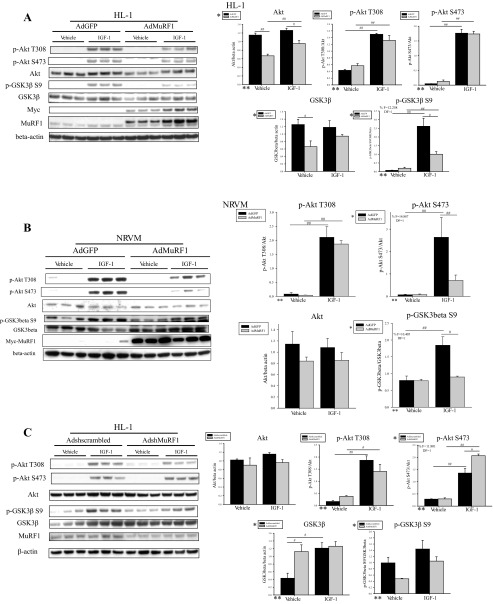
Fig. 3.
MuRF1 inhibits protein expression of Akt and GSK-3β in IGF-I-stimulated HL-1 and NRVM cardiomyocytes. A: HL-1 cells were transduced with AdMuRF1 or control AdGFP at MOI of 25 for 24 h in serum-free DMEM to increase MuRF1 expression, followed by treatment with 10 nM IGF-I for 30 min. Immunoblots using whole cell lysates from 3 independent experiments are shown for p-Akt Ser473, p-Akt Thr308, Akt, GSK-3β, and p-GSK-3β Ser9. Primary antibody against myc was used to assess adenovirus-dependent expression of myc-MuRF1, and immunoblot for MuRF1 was done to determine endogenous protein levels. β-Actin was used as a loading control. Densitometry analysis of Akt, p-Akt Ser473, p-Akt Thr308, GSK-3β, and p-GSK-3β Ser9 is shown for vehicle- or IGF-I-treated cardiomyocytes transduced with either AdGFP or AdMuRF1. Total protein levels were normalized to β-actin, and phosphorylated protein levels were normalized first to β-actin and then to total protein levels. B: NRVM were transduced with AdMuRF1 or control AdGFP at MOI of 25 for 24 h in serum-free M199 to increase MuRF1 expression, followed by treatment with 10 nM IGF-I for 30 min. Immunoblots using whole cell lysates from 3 independent experiments are shown for p-Akt Ser473, p-Akt Thr308, Akt, GSK-3β, and p-GSK-3β Ser9. Primary antibody against myc was used to assess adenovirus-dependent expression of myc-MuRF1. β-Actin was used as a loading control. Densitometry analysis of Akt, p-Akt Ser473, p-Akt Thr308, and p-GSK-3β Ser9 is shown for vehicle- or IGF-I-treated cardiomyocytes transduced with either AdGFP or AdMuRF1. Total protein levels were normalized to β-actin, and phosphorylated protein levels were normalized first to β-actin and then to total protein levels. C: HL-1 cardiomyocytes were transduced with either AdshMuRF1 or control Adshscrambled at MOI of 30 for 48 h in serum-free DMEM, followed by treatment with 10 nM IGF-I for 30 min. Akt and GSK-3β expression and phosphorylation in whole cell lysates from 3 independent experiments were assessed by immunoblot using primary antibodies raised against total Akt or GSK-3β and p-Akt Ser473, p-Akt Thr308, and p-GSK-3β Ser9, as indicated. Primary antibody against MuRF1 was used to confirm knockdown. β-Actin was used as a loading control. Densitometry analysis of Akt, p-Akt Ser473, p-Akt Thr308, GSK-3β, and p-GSK-3β Ser9 is shown for vehicle- or IGF-I-treated cardiomyocytes transduced with either Adshscrambled or AdshMuRF1. Densitometry analysis is represented as means ± SE. A 2-way ANOVA test was used to determine statistical significance. *Significance on level of adenovirus group; **significance on level of treatment group; %significant interaction between adenovirus and treatment groups. The F statistic and DF were reported when dependence between groups was found to be a significant source of variation. #P < 0.05 and ##P < 0.001, significance between groups as determined using a pairwise posttest.

