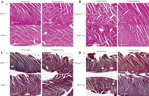
Fig. 8.

High-power histological analysis of MuRF−/−, MuRF1 Tg+, and sibling wild-type control hearts. A and B: hematoxylin and eosin staining of representative cardiac sections from MuRF1−/− (A) and MuRF1 Tg+ mice (B) after 5 wk of sham or running challenge. C and D: Masson's trichrome staining of representative cardiac sections from MuRF1−/− (C) and MuRF1 Tg+ mice (D) after 5 wk of sham or running challenge.
